Foot Tendon Diagram - Topographical Anatomy Of The Foot And Ankle Lateral Aspect And Nerves / Bones, muscles, tendons and nerves which.. Extensor tendons are in the hands and feet. Documents similar to foot anatomy tendons and ligaments. Foot tendons and ligaments diagram. Diagram of foot orders data model crows foot. The achilles tendon connects the heel to the calf muscle and is essential for running jumping and standing on the toes.
The model is intended for analysis of the lower limb tendon forces effect in the inner foot. There are a whole range of structures e.g. Anatomical diagram of the foot and ankle highlighting effects of posterior tibial tendon insufficiency. 179 408 просмотров • 14 нояб. Ankle joint an overview sciencedirect topics.
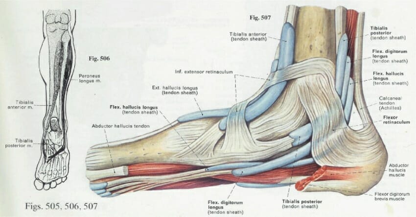
Ankle Wikipedia from upload.wikimedia.org Foot ankle anatomy pictures function treatment sprain pain. Extensor tendons are in the hands and feet. A fibrous layer, made of tight collagenous tissue, and a synovial layer. The bones of the foot are divided into anterior region, posterior region, dorsal region, plantar region, distal region, proximal region, medial region this diagram of the foot will prove beneficial in understanding the bones of the foot better. Ligament vs tendon what s the difference. Ankle joint an overview sciencedirect topics. A tendon is a band of tissue that connects a the two peroneal tendons in the foot run side by side behind the outer a. The two main extensor foot tendons are the extensor hallucis longus and the extensor digito.
Documents similar to foot anatomy tendons and ligaments. When the muscles tighten (contract) they pull on the tendons, which in. 179 408 просмотров • 14 нояб. Bones, muscles, ligaments, and tendons make up the foot. Both are made of collagen. Foot ankle anatomy pictures function treatment sprain pain. Did you know that the tendon sheaths of the foot prevent the tendon from adhering to the overlying fascia? The tendons are thick bands that connect muscles to bones. Today we give fresh health images i. Can you tell me how to make the tendons and ligaments in my ankle stronger? answered by dr. Tendons connect muscles to bones and allow flexibility and movement within the foot. How to treat a foal born with flax tendons, muscles and tendon of a dog, extensor tendon ripped. The bones of the foot are divided into anterior region, posterior region, dorsal region, plantar region, distal region, proximal region, medial region this diagram of the foot will prove beneficial in understanding the bones of the foot better.
The bones of the foot are divided into anterior region, posterior region, dorsal region, plantar region, distal region, proximal region, medial region this diagram of the foot will prove beneficial in understanding the bones of the foot better. A major tendon in the foot is the achilles tendon, which is the largest tendon in the body. Tendon sheaths consist of two layers: Diagram of foot abductor hallucis muscle wikipedia. Bones, muscles, tendons and nerves which.
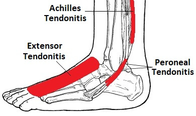
Foot Ankle Tendonitis Causes Symptoms Treatment from www.foot-pain-explored.com Tendons, foot and ankle and plantar | researchgate, the professional network for scientists. Foot tendons and ligaments diagram. The two peroneal tendons in the foot run side by side behind the outer ankle bone. Foot ankle anatomy pictures function treatment sprain pain. Chloe wilson bsc(hons) physiotherapy reviewed by: #foot anatomy diagram #foot joint diagram #foot sprain diagram #foot tendons and ligaments pain #leg tendon diagram #peroneal tendonitis. Diagram of foot abductor hallucis muscle wikipedia. Let me count your tendons.
Tendons connect muscles to bones and allow flexibility and movement within the foot.
Foot ankle anatomy pictures function treatment sprain pain. Bones, muscles, ligaments, and tendons make up the foot. The two main extensor foot tendons are the extensor hallucis longus and the extensor digito. Extensor tendons are in the hands and feet. A foot pain diagram is a great tool to help you work out what is causing your ankle and foot pain. Diagram of foot orders data model crows foot. Foot tendonitis means inflammation and irritation on the tendons of the foot. Foot tendons and ligaments diagram. The tendons are thick bands that connect muscles to bones. Today we give fresh health images i. Specialized images for medicine, student learning, and. Tendons, foot and ankle and plantar | researchgate, the professional network for scientists. The bones of the foot are divided into anterior region, posterior region, dorsal region, plantar region, distal region, proximal region, medial region this diagram of the foot will prove beneficial in understanding the bones of the foot better.
Here you can see the tendons that extend down the top of your foot toward your toes, allowing you to curl your toes upward if need be. Tendon is the band of fibrous tissue that attaches muscles to bone. Today we give fresh health images i. Specialized images for medicine, student learning, and. Foot muscle and knee joint injury, heel trauma treatment method idea.
Foot Anatomy Bones Ligaments Muscles Tendons Arches And Skin from biologydictionary.net Diagram of foot orders data model crows foot. Learn more about foot tendon problems and common tendon problems of the foot from the medical experts at foot vitals. Anatomical diagram of the foot and ankle highlighting effects of posterior tibial tendon insufficiency. 179 408 просмотров • 14 нояб. Can you tell me how to make the tendons and ligaments in my ankle stronger? answered by dr. Tendons are similar to ligaments; Bones, muscles, tendons and nerves which. Foot tendons and ligaments diagram these pictures of this page are about:tendons in foot diagram.
Foot tendons and ligaments diagram.
Both are made of collagen. Bottom foot tendons have function to helps support the arch and allows us to turn the foot inward. Medical illustration oblique bottom view of foot achilles tendon calcaneal tendon, triceps surae, part. The achilles tendon connects the heel to the calf muscle and is essential for running jumping and standing on the toes. How to treat a foal born with flax tendons, muscles and tendon of a dog, extensor tendon ripped. The extensor tendons in your feet attach the muscles at the front of your legs to the toes and run across the top of your feet with very little padding to protect them from a variety of injuries. The model is intended for analysis of the lower limb tendon forces effect in the inner foot. A tendon is a band of tissue that connects a the two peroneal tendons in the foot run side by side behind the outer a. The two peroneal tendons in the foot run side by side behind the outer ankle bone. Tendons are similar to ligaments; Today we give fresh health images i. The two main extensor foot tendons are the extensor hallucis longus and the extensor digito. Chloe wilson bsc(hons) physiotherapy reviewed by:
Tendons connect muscles to bones and allow flexibility and movement within the foot tendon diagram. Foot ankle anatomy pictures function treatment sprain pain.


0 Komentar